The Discovery of the Cell and the Invention of the Microscope
Early Discoveries Through Microscopy
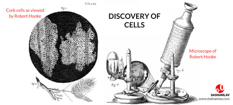
Robert Hooke was an English scientist living in the 1600s. Using one of the earliest microscopes, he viewed a thin slice of cork and saw tiny compartments that resembled monk’s living quarters. He coined the term “cell” to describe these structures in his 1665 book Micrographia. At around the same time, Dutch scientist Anton van Leeuwenhoek independently designed his own improved microscope and began his observations. He is considered the “father of microbiology” for being the first to observe and describe single-celled microorganisms like bacteria. Both men’s work in the late 1600s marked the foundation of cell biology.

The First Microscopes and Their Limitations
Early microscopes were primitive compound microscopes that used multiple lenses to magnify. They had strict limitations compared to modern instruments. Hooke and Leeuwenhoek developed some of the earliest unaided single-lens microscopes, which provided better magnification and resolution than compound models. However, viewfields were very narrow and illumination was poor. Colors were also difficult to distinguish. Nevertheless, these basic microscopes allowed the observers to make their groundbreaking discoveries of cells and microscopic life.
Advances in Lens Design and Construction
In the 1700s and 1800s, optical glass technologies advanced microscope lenses. Skilled craftsmen like Johnston, Dolland, and Smith pushed the boundaries of achievable magnification and image clarity. Compound microscopes added more lens combinations and became the standard design. Key innovations included achromatic lens pairs that reduced chromatic aberration, allowing colored specimens to be observed with finer details. Improved mechanical stages also enhanced usability. By the Victorian era, advanced compound microscopes revealed intricate subcellular structures like organelles. The era of modern cell biology had truly begun.
Illumination Strategies for Microscopy
One thing Hooke and Leeuwenhoek lacked was effective microscope illumination. Early observers used ambient light or light from a nearby window. This made viewing faint specimens quite difficult and limited magnification. In the 1800s, electric lighting was applied to microscopy. Modern incarnations include Köhler illumination setups optimized for contrast and resolution. Darkfield techniques also emerged, allowing structures to be seen by light passing around rather than through them. Brightfield, phase contrast, and fluorescence modes further empowered researchers to visualize even the most delicate intracellular features. Combined with advances in staining techniques, illumination tools are critical to modern microscopic analysis.
Digitization and Computer-Assisted Microscopy
In recent decades, digital imaging sensors and computing power have transformed light microscopy. Video capture allows live dynamic processes to be recorded and analyzed frame-by-frame. Multi-dimensional datasets can be collected through techniques like fluorescent recovery after photobleaching (FRAP) and photoactivation localization microscopy (PALM). Sophisticated image processing corrects artifacts and extracts quantitative data from images. Software also automates tasks like focusing, mosaicking, and 3D reconstruction. Perhaps most remarkably, super-resolution methods break the diffraction limit, revealing previously invisible nanoscale structures. These digital and computational innovations continue pushing the boundaries of what can be seen through the lens.
The Continued Evolution of Light Microscopy
After Hooke, Leeuwenhoek, and centuries of relentless refinement by optical scientists, light microscopes today are remarkably powerful investigative tools. From simple single-lens microscopes to digital microscopy workstations, the technology has evolved enormously. Modern microscopists can choose from diverse modalities optimized for applications like live-cell imaging, pathology diagnostics, materials inspection, and more. The door is also open to emerging techniques like lattice light-sheet microscopy that may reveal even finer structural and dynamic secrets of the microscopic world. Looking ahead, further progress in optical designs, biomarkers, genetic engineering, detectors, and image analysis will undoubtedly continue enhancing our understanding of cells and cellular processes at the highest resolutions. The possibilities to advance biology and medicine through the microscope seem limitless.